Long Bone Diagram Hyaline Cartilage : Structure Of Bone Gross Anatomy Of A Long Bone Microscopic Anatomy Ppt Video Online Download - Hyaline cartilage is a type of connective tissue found in areas such as the nose, ears, and trachea of the human body.
Long Bone Diagram Hyaline Cartilage : Structure Of Bone Gross Anatomy Of A Long Bone Microscopic Anatomy Ppt Video Online Download - Hyaline cartilage is a type of connective tissue found in areas such as the nose, ears, and trachea of the human body.. (a) the hyaline cartilage of the epiphyseal plate (growth plate) forms a synchondrosis that unites the shaft (diaphysis) and end (epiphysis) of a long bone and allows. Cartilage cells (chondrocytes) secrete the fibers and ground substance that make up the cartilage matrix. The epiphyseal plate is a hyaline cartilage plate where growth occurs in children and adolescents, located in the metaphysis at each end of a long bone. At cartilaginous joints, bones are united by hyaline cartilage to form a synchondrosis or by fibrocartilage to form a symphysis. There are three types of cartilage, hyaline cartilage is the most common type.
Cartilage and bone are specialized connective tissues that provide support to other tissues and organs. During embryonic development, hyaline cartilage serves as temporary cartilage models that are essential precursors to the formation of most of the axial and appendicular skeleton. Part 1 in a 7 part lecture on bone and cartilage histology in a flipped human anatomy course taught by wendy riggs. Cartilage occurs where flexibility is required, while bone resists deformation. Articular cartilage is hyaline cartilage that is found on the articular surfaces of bone, which is where bones meet and form joints.
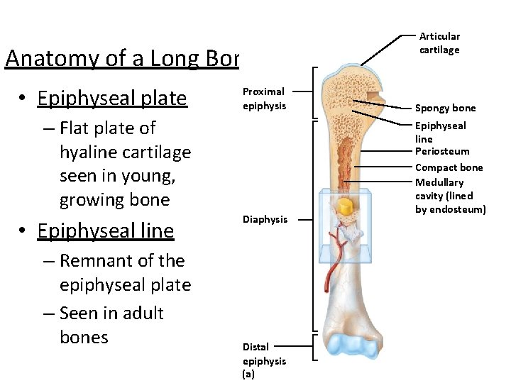
Blank Diagram Of A Long Bone Label The Parts Of A Long Bone The Metaphysis Is The Wide Portion Of A Long Bone Between The Epiphysis And The Reyna Nottingham from i1.wp.com The space in the matrix occupied by a chondrocyte is. The epiphyseal plate is a hyaline cartilage plate where growth occurs in children and adolescents, located in the metaphysis at each end of a long bone. End of the bone located farthest away from the midline 8. Bars of hyaline cartilage (the costal cartilages) connect ribs to sternum. Cartilaginous joints connected by hyaline cartilage are termed synchondroses. The hyaline cartilage occurs in the nasal septum, trachea, ends of the growing bones, and in between the ribs and the sternum. Fibrocartilage attaches bones to other bones and provides restricted mobility to the joints. Cartilage, connective tissue forming the mammalian embryonic skeleton prior to bone formation and persisting in parts of the human skeleton into three main types of cartilage can be distinguished.
Cartilage cells (chondrocytes) secrete the fibers and ground substance that make up the cartilage matrix.
Covers ends of long bones. Fibrocartilage attaches bones to other bones and provides restricted mobility to the joints. (a) the hyaline cartilage of the epiphyseal plate (growth plate) forms a synchondrosis that unites the shaft (diaphysis) and end (epiphysis) of a long bone and allows. …unlike other long bones of the skeleton, vertebral body epiphyses never ossify, and after the end of the growth period of life they are reduced into thin the entire thing is called intervertebral symphysis. Cartilage cells (chondrocytes) secrete the fibers and ground substance that make up the cartilage matrix. | (a) … перевести эту страницу. Watch the whole lecture (all 7. This is known as articular cartilage. This article will focus on important features of hyaline cartilage, namely its matrix, chondrocytes, and perichondrium. Articular cartilage is hyaline cartilage that is found on the articular surfaces of bone, which is where bones meet and form joints. Hyaline cartilage and bone replacement. Hyaline cartilage that covers ends of bones in synovial joi… Cartilaginous joints are a type of joint where the bones are entirely joined by cartilage, either hyaline cartilage or fibrocartilage.
It is also most commonly found in the ribs, nose, larynx, and trachea. Watch the whole lecture (all 7. Further, cartilage is avascular, whereas bone is well permeated there are three types of cartilage which traditionally have been distinguished on the basis of matrix characteristics. Cartilage occurs where flexibility is required, while bone resists deformation. So, where is hyaline cartilage found?
The Skeletal System 1 The Skeletal System Parts from slidetodoc.com The tumor matrix had a distinctive appearance of homogeneous high signal intensity in a defined lobular configuration on images of all hyaline cartilage lesions obtained with a long. The white fibrous cartilage have matrix of densely packed white collagen fibres. Bars of hyaline cartilage (the costal cartilages) connect ribs to sternum. Glycosaminoglycans, chiefly chondroitin sulfate, are contained. This is known as articular cartilage. Covers ends of long bones. Watch the whole lecture (all 7. Related online courses on physioplus.
Cartilaginous joints connected by hyaline cartilage are termed synchondroses.
Cartilage cells (chondrocytes) secrete the fibers and ground substance that make up the cartilage matrix. Hyaline cartilage is the most widespread and is the type that makes up the embryonic skeleton. • by the property of cartilage tissue (hyaline or fibrous), there are End of the bone located farthest away from the midline 8. The long bones of the body can maintain their increase in length as the body grows due to the interstitial growth of hyaline cartilage in the epiphyseal plate. Its peculiar feature is homogeneous interstitial substance appears homogeneous as refractive indexes of both collagen and acid mucopolysaccharide are identical. This article will focus on important features of hyaline cartilage, namely its matrix, chondrocytes, and perichondrium. The hyaline cartilage occurs in the nasal septum, trachea, ends of the growing bones, and in between the ribs and the sternum. The epiphyseal plate is a hyaline cartilage plate where growth occurs in children and adolescents, located in the metaphysis at each end of a long bone. The space in the matrix occupied by a chondrocyte is. The white fibrous cartilage have matrix of densely packed white collagen fibres. At cartilaginous joints, bones are united by hyaline cartilage to form a synchondrosis or by fibrocartilage to form a symphysis. During embryonic development, hyaline cartilage serves as temporary cartilage models that are essential precursors to the formation of most of the axial and appendicular skeleton.
Elastic cartilage has abundant elastic fibers in addition to collagen, making the matrix much more elastic than hyaline cartilage. Hyaline cartilage is the most widespread and is the type that makes up the embryonic skeleton. Three types of cartilage are recognized based on differences in fiber composition: • by the property of cartilage tissue (hyaline or fibrous), there are These findings suggest that regeneration of meniscal cartilage through a collagen scaffold is possible.
352 Hyaline Cartilage Stock Photos Pictures Royalty Free Images Istock from media.istockphoto.com Cartilage is distinguishable from bone on the basis of matrix hardness and density. Part 1 in a 7 part lecture on bone and cartilage histology in a flipped human anatomy course taught by wendy riggs. Hyaline cartilage covers bone surfaces at synovial joints. Hyaline cartilage provides mechanical support for the respiratory tree, nose, articular surfaces, and developing bones. Hyaline cartilage (some articular cartilage), fibrocartilage, and fibrous tissue. Articular cartilage is hyaline cartilage that is found on the articular surfaces of bone, which is where bones meet and form joints. Gags are essentially long polysaccharides made of amino sugars that attract sodium and potassium ions. Cartilage and bone are specialized connective tissues that provide support to other tissues and organs.
This is known as articular cartilage.
During embryonic development, the hyaline cartilage will form the skeleton of the. Cartilage and bone are specialized connective tissues that provide support to other tissues and organs. Tute was created with a zcc interfacing them. Cartilage, connective tissue forming the mammalian embryonic skeleton prior to bone formation and persisting in parts of the human skeleton into three main types of cartilage can be distinguished. Part 1 in a 7 part lecture on bone and cartilage histology in a flipped human anatomy course taught by wendy riggs. The long bones of the body can maintain their increase in length as the body grows due to the interstitial growth of hyaline cartilage in the epiphyseal plate. The white fibrous cartilage have matrix of densely packed white collagen fibres. Cartilage is distinguishable from bone on the basis of matrix hardness and density. Forms most of embryonic skeleton. The epiphyseal plate is a hyaline cartilage plate where growth occurs in children and adolescents, located in the metaphysis at each end of a long bone. Elastic cartilage has abundant elastic fibers in addition to collagen, making the matrix much more elastic than hyaline cartilage. Hyaline cartilage is vulnerable because it has no blood supply; Glycosaminoglycans, chiefly chondroitin sulfate, are contained.
Cartilaginous joints are a type of joint where the bones are entirely joined by cartilage, either hyaline cartilage or fibrocartilage long bone diagram. At cartilaginous joints, bones are united by hyaline cartilage to form a synchondrosis or by fibrocartilage to form a symphysis.


0 Komentar